SIXPAD的效果驗證
將EMS的正確理論和效果,
廣泛正確地傳達到世間的努力
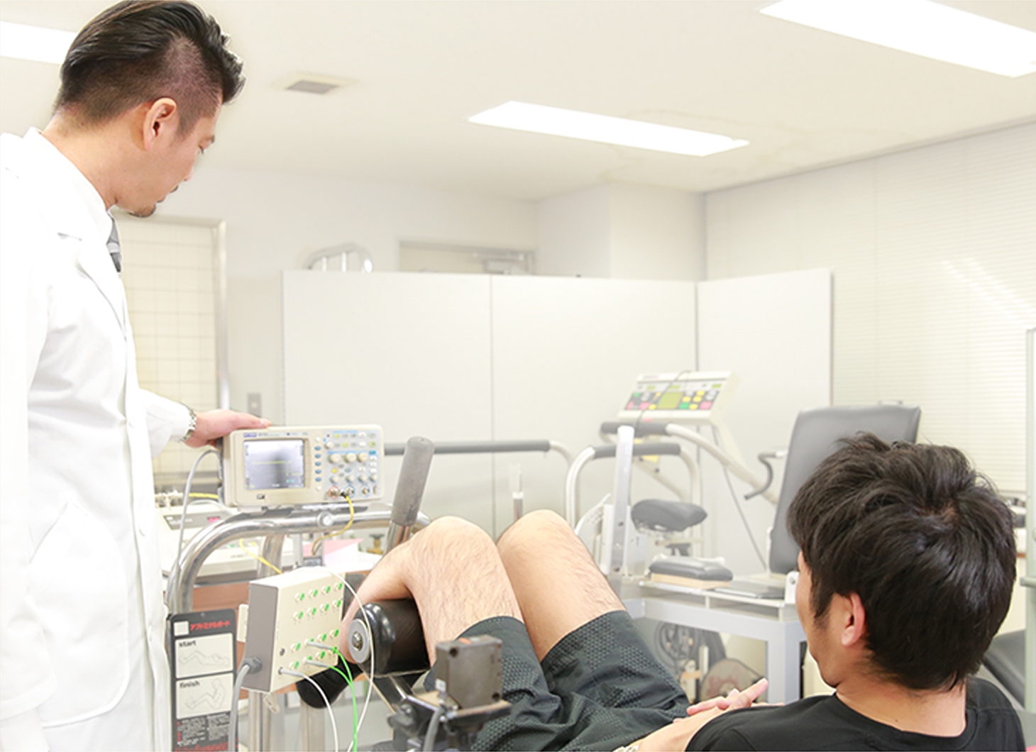
SIXPAD的人體效果驗證
與中京大學國際教養學系渡邊航平副教授共同進行驗證,SIXPAD鍛鍊(肌肉電刺激)所產生的肌肉收縮是否能引起生理學上的肌肉疲勞。
Training Suit的
效果驗證
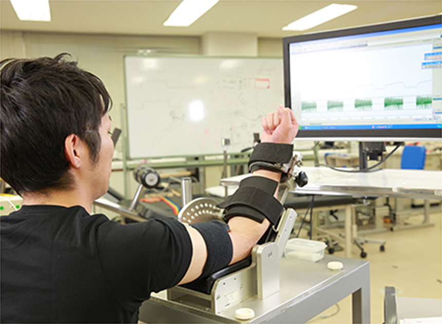
實際驗證肌肉活動水平的提高。
驗證項目
在穿著訓練褲與未穿著訓練褲時進行相同的動作(膝關節從180度彎曲到90度),比較大腿膕繩肌的活動電位。
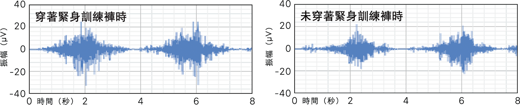
結果發現,穿著訓練褲時,肌電圖振幅值增加了約30%*,與未穿著訓練褲時相比,證實可產生更多的神經肌活動。這可以解釋為,即使是相同的運動,也能反映出大腿膕繩肌在穿著訓練褲時會試圖進行更強烈的運動。
*受試者10名(31 歲~ 50 歲男性)在未穿著訓練褲與穿著訓練褲時的肌電圖振幅值的平均值比較(第三方機關調查)
大腿膕繩肌表面肌電圖波形的範例
協助說明實驗數據:渡邊航平 中京大學 國際教養學部 副教授
中京大學運動科學系渡邊航平教授
- 2010年取得名古屋大學研究所教育發達科學研究科博士學位
- 2010年擔任京都大學研究所人類和環境學研究科特別研究員
- 2012年起擔任中京大學國際教養學系副教授
- 2020年中京學教养教育研究院教授
- 2021年中京學運動科學系教授
隸屬於日本體育學會、日本體力醫學會、日本生物力學會、日本運動生理學會、日本電氣生理運動學會、美國運動醫學會(ACSM)、國際電氣生理運動學會。
專業領域為運動生理學、生物力學,針對運動時身體各機能的變化和適應,將焦點放在「骨骼肌和支配骨骼肌的運動神經」,以人類為對象進行研究。